FIGURE

FIGURE 3

ID
ZDB-FIG-250214-35
Publication
García-Castro et al., 2025 - Phenogenetics of cortical granule dynamics during zebrafish oocyte-to-embryo transition
Other Figures
All Figure Page
Back to All Figure Page
FIGURE 3

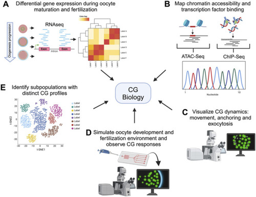
Comprehensive techniques for studying cortical granule biology. Holistic view of the tools available to study the processes involved in CG biology. The figure illustrates various advanced techniques used to examine different aspects of CG biology. Each technique provides unique information into the CG lifecycle, from biosynthesis to exocytosis. (A) Transcriptomics analyzes differential gene expression during oocyte maturation and fertilization, offering insights into the molecular regulation of CG biology. (B) ChIP-Seq and ATAC-Seq map chromatin accessibility and transcription factor binding sites, elucidating the genetic regulation of CG biology. (C) High-resolution live-cell microscopy and Super-resolution imaging allows to visualize CG dynamics, including movement, anchoring, and exocytosis in real-time. (D) Microfluidic devices simulate the oogenesis and fertilization environment, allowing observation of CG responses in controlled conditions. Synthetic biology enables the design of constructs to mimic or disrupt CG pathways, facilitating the study of their functional roles. (E) Single-cell sequencing identifies subpopulations of cells with distinct CG profiles, highlighting cellular diversity.

Expression Data

Expression Detail
Antibody Labeling
Phenotype Data

Phenotype Detail
Acknowledgments
This image is the copyrighted work of the attributed author or publisher, and ZFIN has permission only to display this image to its users. Additional permissions should be obtained from the applicable author or publisher of the image. Full text @ Front Cell Dev Biol