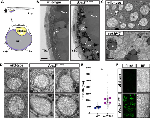
Cytoplasmic lipid droplets and swollen, electron-dense ER are present in the yolk syncytial layer of dgat2 mutants.A, depiction of the cross-sectional view of a 4 dpf zebrafish illustrating the yolk syncytial layer (YSL) surrounding the yolk mass. The dashed box indicates the location of the images in panel B. B, representative transmission electron micrographs of the yolk and YSL from wild-type and dgat2sa13945 mutants; dashed lines delineate the YSL region, n = nucleus, YP = yolk platelet, LD and ∗ = lipid droplet, scale = 10 μm. C, higher magnification images of the YSL region. Arrows indicate the endoplasmic reticulum which often encircles the mitochondria (m) in both wild-type and dgat2sa13945 mutants; scale = 1 μm. D, examples of ER morphology in wild-type and dgat2sa13945 embryos; scale = 0.5 μm. E, quantification of ER width, N = 4 fish per genotype (30–40 measurements per fish from 3 to 4 images, mean of each fish is shown by the blue and magenta points); bars indicate overall mean ± SD, unpaired t test, ∗∗p = 0.0022. F, representative confocal z-sections of the YSL in a wild-type fish and dgat2sa13945 mutant carrying the Fus(EGFP-plin2)/+ reporter; scale = 20 μm, 3 dpf, n = 18 dgat2sa13945 & n = 15 WT or dgat2sa13945/+ fish from two independent experiments.
|
