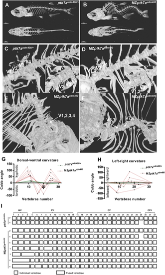
MZptk7audm400 mutants display a severe 3D spinal curvature and CVMs restricted to the anterior part of the spine. (A,B) Representative μCT images of ptk7audm400/+ (A) and MZptk7audm400 (B) 3 months postfertilization adult females (lateral views, top; dorsal views, bottom). MZptk7audm400 adults display severe 3D curvature defects. (C) Representative μCT images of ptk7audm400/+ adult fish showing a lateral view of a normally segmented anterior part of the spine (dorsal view of V1-V4 is shown in the inset). (D) Representative μCT images of MZptk7audm400 adult fish showing the most common CVM defect (fusion of V1 and V2) from lateral and dorsal view (inset). (E,F) Examples of lateral views of other severe CVMs detected in MZptk7audm400 adult fish, including fused V1-V4 (E) and mis-segmentation of V6-V8 (F). (G,H) Quantification of curve severity, direction, and position along dorsal-ventral (G) and left-right (H) axes. (I) Diagram of the CVMs detected in MZptk7audm400 (n=8) compared with ptk7audm400/+ (n=8). All CVMs were restricted to the anterior part of the spine spanning V1 to V9. CFV, caudal fin vertebrae; CV, caudal vertebrae; PV, precaudal vertebrae; WA, Weberian vertebrae. Scale bars: 2 mm.
|
