FIGURE

Fig. 3

ID
ZDB-FIG-241123-4
Publication
Li et al., 2024 - A Huluwa phosphorylation switch regulates embryonic axis induction
Other Figures
All Figure Page
Back to All Figure Page
Fig. 3

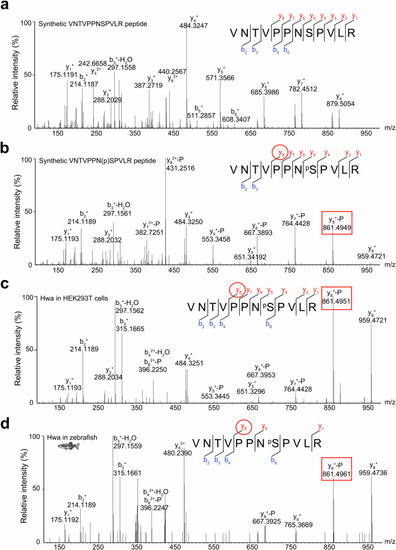
The phosphorylation of Hwa protein at Ser168 was validated by LC-MS/MS.

Synthetic nonphosphorylated (a) and phosphorylated (b) peptides were used as references for the nonphosphorylated and phosphorylated states, respectively. Flag-tagged Hwa proteins purified from HEK293T cells (c) and zebrafish embryos (d) were used for phosphorylation identification, and the MS spectra of fragments containing VNTVPPNSPVLR were compared with that of phosphorylated peptide (b) to ensure phosphorylation at the Ser168 site. The labels “y” and “b” designate the C- and N- terminal peptide fragment ions, respectively, which were produced by collision-induced fragmentation at the peptide bond. The subscripted number (e.g., y8, b2,) represents the number of C- or N-terminal residues in the peptide fragment. The labels “-H2O” and “-P” designate ions with water (H2O) and phosphoric acid (H3PO4) loss, respectively. The red circle indicates the specific collision-induced fragmentation to identify the phosphorylation state and the red rectangle indicates the corresponding peak in the MS profile. Source data are provided as a Source Data file.

Expression Data

Expression Detail
Antibody Labeling
Phenotype Data

Phenotype Detail
Acknowledgments
This image is the copyrighted work of the attributed author or publisher, and ZFIN has permission only to display this image to its users. Additional permissions should be obtained from the applicable author or publisher of the image. Full text @ Nat. Commun.