
FIGURE
Fig. 3
- ID
- ZDB-FIG-240918-48
- Publication
- Albu et al., 2024 - Distinct mechanisms regulate ventricular and atrial chamber wall formation
- Other Figures
- All Figure Page
- Back to All Figure Page
Fig. 3
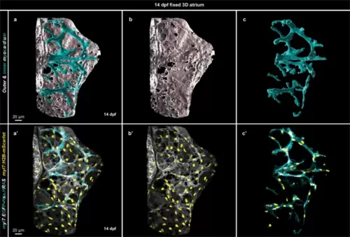
Elongated cardiomyocytes form the inner atrial muscle structures.a–c 3D surface rendering of a 14 dpf fixed atrium; outer layer myocardium shown in white and inner layer myocardium in cyan. a’–c’ 3D confocal image of the atrium from which the surface rendering was created; CM membranes shown in white for outer layer CMs and in cyan for inner layer CMs; all CM nuclei are shown in yellow. |
Expression Data
Expression Detail
Antibody Labeling
Phenotype Data
Phenotype Detail
Acknowledgments
This image is the copyrighted work of the attributed author or publisher, and
ZFIN has permission only to display this image to its users.
Additional permissions should be obtained from the applicable author or publisher of the image.
Full text @ Nat. Commun.