|
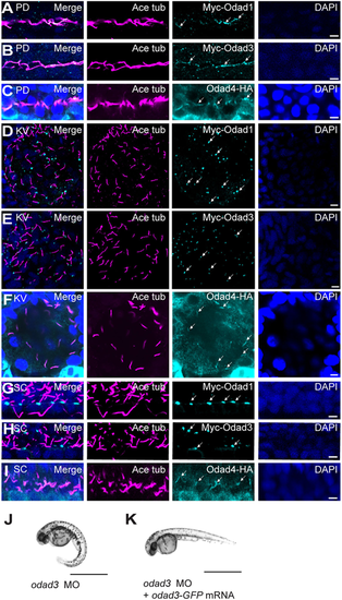
Differential localisation of Odad proteins to zebrafish motile cilia axonemes and basal bodies. (A-C) Odad1, Odad3 and Odad4 localised along axonemes of (9+2) cilia in zebrafish pronephric duct (PD) at 24 hpf. (D,E) Odad1 and Odad3 localised to KV cilia base at the 10-somite stage. (F) Odad4 localised along axonemes of KV cilia. (G,H) Odad1 and Odad3 localised at the spinal canal (SC) cilia base at 24 hpf. (I) Odad4 localised along axonemes of SC cilia at 24 hpf. Odad proteins were detected with anti-Myc antibody (for Odad1 and Odad3; cyan, arrows) or anti-HA antibody (for Odad4; cyan, arrows), cilia with anti-acetylated tubulin antibody (magenta) and nuclei with DAPI (blue). (J) An odad3 morphant (MO) (48 hpf), showing a ventrally curved body axis. (K) An odad3 morphant (MO) (48 hpf), with rescue of axial curvature on co-injection of odad3-gfp mRNA. Data represent three technical replicates with n=75 per replicate. Scale bars: 5 µm (A-I); 1 mm (J,K).
|