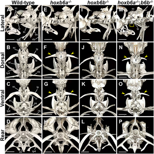
Zebrafish hoxb6a−/−;hoxb6b−/− mutants exhibit shortening of the lateral process in the second vertebra. (A-P) The anterior vertebrae were analyzed by micro-CT scan in wild-type (A-D, n=4), hoxb6a−/− (E-H, n=2), hoxb6b−/− (I-L, n=2) and hoxb6a−/−;hoxb6b−/− (M-P, n=2) fish. Adult zebrafish of the same genotype subjected to micro-CT scanning showed similar morphology; therefore, representative images are shown. Fish were obtained from multiple intercrosses between hoxb6a+/−;hoxb6b+/− fish, and genotype analysis of the surviving juveniles is shown in Table S1B. The numbering indicates the position of each vertebra from the first vertebra. The arrowheads indicate the reduced lateral process on the second centrum in hoxb6a−/− and hoxb6a−/−;hoxb6b−/− mutants. The brackets signify the vertebra with both scaphium and lateral process. Scale bars: 1 mm. For details, micro-CT scan 3D movies are provided in Movies 5,6,7. A summary of the phenotypes of these mutants is shown in Table S2. Digital dissection of each vertebra in hoxb6a−/−;hoxb6b−/− is shown in Fig. S3. ic, intercalarium; lp, lateral process; os, os suspensorium; sc, scaphium; tp4, transverse process of vertebra 4; tri, tripus.
|
