
Fig. 7
- ID
- ZDB-FIG-240701-31
- Publication
- Pollitt et al., 2024 - Llgl1 mediates timely epicardial emergence and establishment of an apical laminin sheath around the trabeculating cardiac ventricle
- Other Figures
- All Figure Page
- Back to All Figure Page
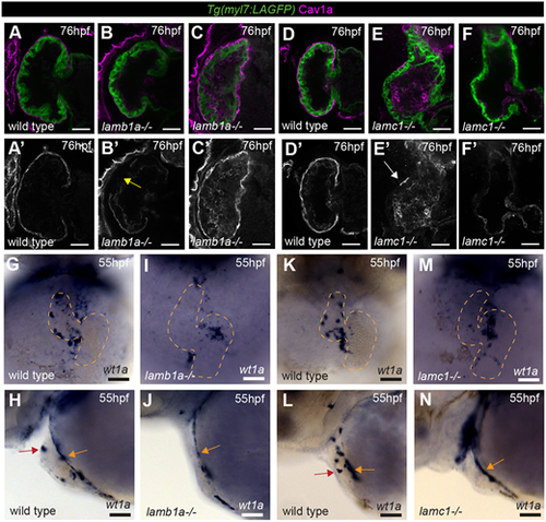
Laminin promotes epicardial emergence and ventricular colonisation. (A-F) Confocal z-slices through the ventricle of Tg(myl7:LifeAct-GFP) transgenic embryos visualising the myocardium (green) and anti-Cav1a antibody (magenta), highlighting the epicardium at 76 hpf in wild-type siblings (A,D), lamb1a mutants (B,C) and lamc1 mutants (E,F). A′-F′ show Cav1a staining only. Scale bars: 25 µm. Wild-type embryos show full epicardial coverage at 76 hpf (A, n=12/12; D, n=11/11), whereas lamb1a and lamc1 mutants show defects in epicardial coverage. The majority of lamb1a mutants have partial epicardial coverage (B′, yellow arrow, n=10/13), with a small number exhibiting full epicardial coverage (C, n=3/13). lamc1 mutants have more profound epicardial defects, with the majority having no epicardial cells attached at 76 hpf (F, n=6/9), and a small proportion having only a few epicardial cells attached (E′, white arrow, n=3/9). (G-N) mRNA in situ hybridisation analysis of wt1a expression at 55 hpf in wild-type embryos (G,H,K,L), lamb1a mutants (I,J) and lamc1 mutants (M,N). G,I,K,M show the ventral view, dashed line outlines the heart; H,J,L,N show the lateral view. Scale bars: 50 µm. Similar to wild-type siblings (H, n=10; J, n=12), both laminin mutants have wt1a expression in proepicardial cells on the dorsal pericardium (L, n=11; N, n=12; orange arrows). Wild-type embryos also have epicardial cells attached to the ventral ventricular wall (H,L; red arrows) but both lamb1a and lamc1 mutants show little attachment of epicardial cells to the ventral ventricle at 55 hpf (J,N). |