Fig. 6
- ID
- ZDB-FIG-240620-56
- Publication
- Zhang et al., 2024 - Sevoflurane postconditioning ameliorates cerebral hypoxia/reoxygenation injury in zebrafish involving the Akt/GSK-3β pathway activation and the microtubule-associated protein 2 promotion
- Other Figures
- All Figure Page
- Back to All Figure Page
|
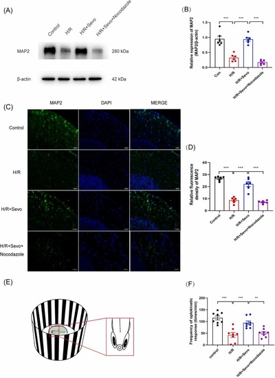
Sevoflurane postconditioning promoted MAP2 expression and zebrafish visual neurobehavioral recovery. (A) Representative western blotting images showing MAP2 protein levels in the control, H/R, H/R + Sevo, and H/R + Sevo + Nocodazole groups. (B) Band intensity analysis showing the ratio of MAP2 to β-actin. (C) Representative images of MAP2 immunofluorescence in the zebrafish midbrain. (D) Relative intensity analysis of MAP2-positive cells in the control, H/R, H/R + Sevo, and H/R + Sevo + Nocodazole groups. (E) The schematic diagram of the experimental setup used for investigating optokinetic response behavior in adult zebrafish. (F) Statistical analysis of the frequency of optokinetic response of zebrafish in the control, H/R, H/R + Sevo, and H/R + Sevo + Nocodazole groups. Six animals in each group were used in western blotting and immunofluorescence staining. Eight animals in each group were used to in optokinetic response behavior. Scale bar = 500 μm. Data are expressed as mean ± S.E.M. The significance levels were ** p < 0.01 and ***p < 0.001, with analyzed by one-way ANOVA followed by Tukey’s multiple comparison test |

