
FIGURE
Fig. 1
- ID
- ZDB-FIG-240528-55
- Publication
- Gölz et al., 2023 - Development of the integrated fish endocrine disruptor test (iFEDT) - Part B: Implementation of thyroid-related endpoints
- Other Figures
- All Figure Page
- Back to All Figure Page
Fig. 1
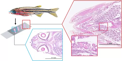
Cutting planes and representative histological sections of eye and thyroid tissues in juvenile zebrafish (Danio rerio). In the region of the gills, isolated thyroid follicles are dispersed close to the ventral aorta. The follicular shape ranges from round to oval, with cuboidal epithelium and uniformly stained colloids. Eye sections are taken at the level of the optical nerve. Sections of 2.5 μm thickness and stained with hematoxylin and eosin G (H&E) |
Expression Data
Expression Detail
Antibody Labeling
Phenotype Data
Phenotype Detail
Acknowledgments
This image is the copyrighted work of the attributed author or publisher, and
ZFIN has permission only to display this image to its users.
Additional permissions should be obtained from the applicable author or publisher of the image.
Full text @ Integr. Environ. Assess. Manag.