|
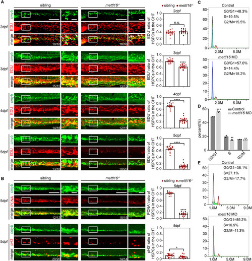
Depletion of Mettl16 inhibits HSPC proliferation through G1/S cell cycle arrest. (A) Double immunostaining of cmyb: EGFP and EDU showing the number of proliferating HSPCs in the CHT of siblings and mettl16 mutants from 2 dpf to 5 dpf. Numbers at the bottom right indicate the number of embryos with similar staining patterns among all embryos examined. n = 3 independent experiments. The white arrowheads indicate proliferating HSPCs. Scale bars, 40 μm. (B) Double immunostaining of cmyb: EGFP and PCNA (upper panels) or pH3 (lower panels) showing the number of proliferating HSPCs in the CHT of siblings and mettl16 mutants at 5 dpf. Numbers at the bottom right indicate the number of embryos with similar staining patterns among all embryos examined. n = 3 independent experiments. Scale bars, 40 μm. (C, D) Flow analysis showing the cell cycle of HSPCs of mettl16-deficient zebrafish at 3 dpf. n ≥ 200 per group, performed with three biological replicates. (E) Flow analysis showing the cell cycle of HSPCs of mettl16-deficient zebrafish at 5 dpf. n ≥ 200 per group, performed with three biological replicates. Data information: In (A, B, D), data were represented as mean ± SEM, * adjusted P < 0.05, ** adjusted P < 0.01, *** adjusted P < 0.001, **** adjusted P < 0.0001, n.s. non-significant, Student’s t-test. Source data are available online for this figure.
|