
Fig. 1
- ID
- ZDB-FIG-240514-54
- Publication
- McLeod et al., 2023 - Specific CaMKIIs mediate convergent extension cell movements in early zebrafish development
- Other Figures
- All Figure Page
- Back to All Figure Page
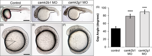
camk2b1 and camk2g1 morphant phenotypes. (A–C). Lateral view of 24hpf zebrafish embryos injected with 3 ng mismatch morpholino oligonucleotide (MO), 3 ng camk2b1 MO, or 1.5 ng camk2g1 MO. Control embryos show evenly spaced somites (arrow) with full yolk extension (asterisk). Camk2b1 morphants exhibit compression of the somites, curvature of the tail and reduced yolk extension. Camk2g1 morphants lack yolk and tail extension and exhibit compression of the somites. Scale bar 0.5 mm. (D–F) Lateral view images at the 10 somite stage (ss) of representative morphant embryos. Body gap angles were derived from the head-yolk-tail angle (dashed lines, D). Scale bar 0.25 mm. (G) Body gap angles of embryos at the 10–11ss were averaged from 34 to 80 embryos per condition across three replicated matched studies. ****p < .0001 compared to control. |