
Fig. 2
- ID
- ZDB-FIG-240425-23
- Publication
- Iglesias Gonzalez et al., 2024 - Perturbed development of calb2b expressing dI6 interneurons and motor neurons underlies locomotor defects observed in calretinin knock-down zebrafish larvae
- Other Figures
- All Figure Page
- Back to All Figure Page
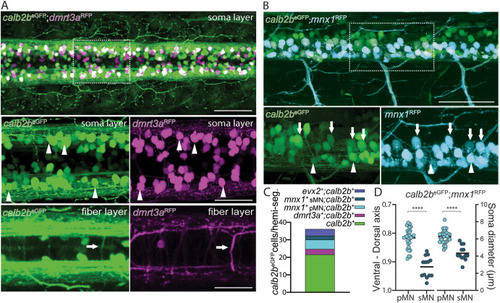
Colocalization of calb2beGFP with dI6-dmrt3a and motor neurons. A) Dorsal view of calb2beGFP; dmrt3aRFP cell bodies and fiber layer at 3 days post fertilization (dpf). Arrowheads indicate overlapping cells and arrows the axon of the CoLo subtype. B) Lateral view of calb2aeGFP; mnx1RFP at 3 dpf. Asterisks mark primary motor neurons and arrowheads mark secondary motor neurons. C) Percentage of calb2beGFP that are colocalized with primary and secondary motor neurons, Evx2 and dI6-dmrt3a interneurons. D) Quantification of calb2aeGFP; mnx1RFP motor neurons by position in the dorso-ventral axis and soma size at 3 dpf. pMN – primary motor neuron, sMN – secondary motor neuron. Scale bar, 50 μm in A, B, C, D and G; 25 μm in zoom boxes in A, B and D. |
Reprinted from Developmental Biology, 508, Iglesias Gonzalez, A.B., Koning, H.K., Tuz-Sasik, M.U., van Osselen, I., Manuel, R., Boije, H., Perturbed development of calb2b expressing dI6 interneurons and motor neurons underlies locomotor defects observed in calretinin knock-down zebrafish larvae, 778777-87, Copyright (2024) with permission from Elsevier. Full text @ Dev. Biol.