
Fig. 7
- ID
- ZDB-FIG-240314-14
- Publication
- Chiang et al., 2023 - The Role of MAPRE2 and Microtubules in Maintaining Normal Ventricular Conduction
- Other Figures
- All Figure Page
- Back to All Figure Page
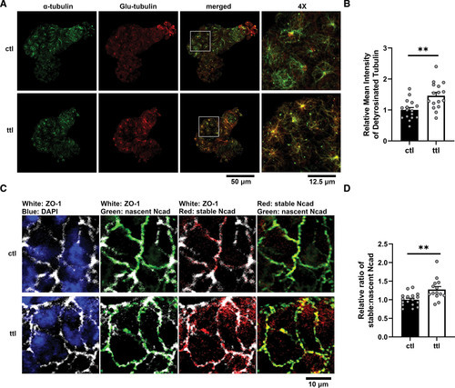
Knockdown of ttl restores fraction of detyrosinated tubulin and stable-to-nascent Ncad (N-cadherin) in mapre2 knockout (KO) hearts. A, Representative immunostaining of hearts from homozygous mapre2 KO larvae injected with control (ctl) vs ttl morpholinos showing a restoration of ventricular detyrosinated tubulin (Glu-tubulin) signal relative to total α-tubulin signal. B, Quantification of ventricular Glu-tubulin signal using α-tubulin signal as a mask (unpaired t test, P=0.0017). C, Immunocytochemistry of hearts from homozygous mapre2 KO larvae on the transgenic background with cdh2 tandem fluorescent timer (tFT). Immunostaining of ZO-1 (zonula occludens-1) was used to mark cell borders. D, Quantification of green fluorescent protein (GFP) and red fluorescent protein (RFP) signals using the ZO-1 signal as mask shows a significant increase in stable-to-nascent Ncad localization at cell borders (unpaired t test, P=0.0028), suggesting a restoration of Ncad balance at adherens junctions. Representative images were chosen based on closeness to group mean and image quality. Each dot represents 1 heart. |