
Fig. 1
- ID
- ZDB-FIG-240304-38
- Publication
- Wu et al., 2023 - Zebrafish ppp1r21 mutant as a model for the study of primary biliary cholangitis
- Other Figures
- All Figure Page
- Back to All Figure Page
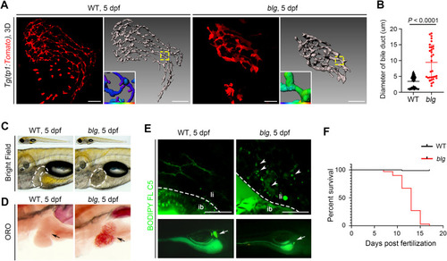
The blg mutant exhibits intrahepatic bile duct anomalies. A: Confocal and 3D reconstruction images of the intrahepatic bile ducts in the WT and blg mutant at 5 dpf under the Tg(tp1:Tomato) transgenic line. The dendrite mean diameter colormaps represent bile duct branches (yellow dotted boxes). The blg mutant showed intrahepatic bile duct anomalies with loss of branches. B: Statistical diagram of intrahepatic bile duct diameter in the WT (n = 40) and blg mutant (n = 13) at 5 dpf. Data are expressed as mean ± SEM, Student's t-test. C: Bright-filed micrographs of the WT and blg mutant at 5 dpf, white dotted outlines indicate the liver area. D: ORO staining of the WT and blg mutant at 5 dpf. Black arrows indicate the liver area. E: BODIPY FL C5 fluorescence staining of the WT (18/18) and blg mutant (13/15) at 5 dpf. The blg mutant presents defective bile flow (arrowheads) and a diminished gallbladder (arrows). F: Survival rate of the WT (n = 80) and blg mutant (n = 70). WT, wild-type; ib, intestinal bulb; li, liver; dpf, days post fertilization. Scale bars, 50 μm (A and E). n = number of embryos with indicated phenotype/total analyzed in each class. |
Reprinted from Journal of genetics and genomics = Yi chuan xue bao, 50(12), Wu, C., Zhang, W., Luo, Y., Cheng, C., Wang, X., Jiang, Y., Li, S., Luo, L., Yang, Y., Zebrafish ppp1r21 mutant as a model for the study of primary biliary cholangitis, 1004-1013, Copyright (2023) with permission from Elsevier. Full text @ J. Genet. Genomics