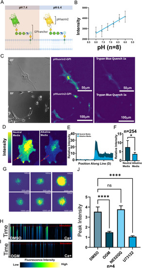
Glioblastoma activates GPR68 by acidifying their extracellular milieu. (A) Schematic representation of the extracellular pH reporter GPI-anchored pHluorin2 (pHluorin2-GPI), which increases in fluorescence intensity upon acidification. (B) Strong correlation between fluorescence intensity and extracellular pH in cells stably expressing pHluorin2-GPI, imaged at 469 nm excitation/525 nm emission. (C) pHluorin2-GPI fluorescence was quenched by the vital dye trypan blue, which is excluded from live cells, confirming that the visualized acidic micro-domains are extracellular. (D) U87 glioma cells expressing pHluorin2-GPI reporter exhibited higher-intensity fluorescence, particularly at cellular protrusions in neutral pH media. Fluorescence was markedly attenuated within 20 s of buffering to pH 8.4 (After), confirming the correlation of fluorescence intensity with low extracellular pH. (E) Quantification of fluorescence intensity along the red line in (A) confirmed a drastic reduction at pH 8.4. (F) The overall cellular intensity of the pHluorin2-GPI signal was reduced by the addition of an alkaline buffer (P < 0.05, n = 6). (G) When grown in spheroids, the extracellular acidification increases over time and becomes more organized (H, I) Kymograph of U87 cells (H) responding to acidification (stimulation) with calcium release. OGM (I) greatly attenuated acid-induced calcium release, in contrast to DMSO vehicle control. (J) Peak calcium responses of U87 cells to acid stimulation in the presence of the GPR68 inhibitor OGM, the GPR4 inhibitor NE52-QQ or the PLC inhibitor U73122 reveals that the response is mediated specifically by the GPR68-PLC pathway, but not by GPR4 (N = 6 OGM, P < 0.0001; NE52QQ, not significant; U73122, P < 0.0001)
|
