
Fig. 6
- ID
- ZDB-FIG-231204-21
- Publication
- Zhang et al., 2023 - Zebrafish MAP2K7 Simultaneously Enhances Host IRF7 Stability and Degrades Spring Viremia of Carp Virus P Protein via Ubiquitination Pathway
- Other Figures
- All Figure Page
- Back to All Figure Page
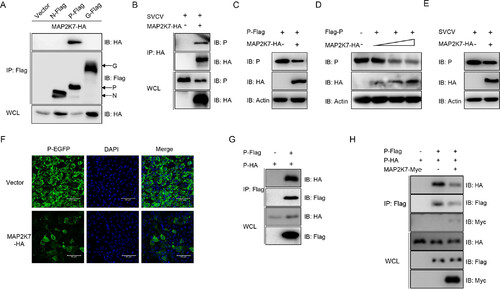
MAP2K7 associates with and degrades the SVCV P protein. (A) EPC cells transfected with the indicated plasmids (5 μg each). After 24 h, cell lysates were immunoprecipitated (IP) with anti-Flag affinity gels. (B) EPC cells seeded in 10 cm2 dishes were transfected with MAP2K7-HA (5 μg). After 24 h, the earlier cotransfected cells were infected with SVCV (MOI, 1); 24 h later, cell lysates were IP with anti-HA affinity gels. (C) EPC cells were transfected with 1 μg P-Flag and 1 μg MAP2K7-HA for 24 h. (D) EPC cells were transfected with P-Flag (1 μg) and MAP2K7-HA (0.25, 0.5, or 1 μg) for 24 h. (E) EPC cells were transfected with MAP2K7-HA. After 24 h, the cells were infected with SVCV (MOI, 1). (F) EPC cells were cotransfected with P-EGPF and vector or MAP2K7-HA for 24 h; then the cells were fixed and subjected to confocal microscopy analysis. Green signals represent the SVCV P protein, and blue staining indicates the nucleus region (original magnification, ×20). Scale bar, 50 μm. (G and H) EPC cells were transfected with 5 μg P-Flag and 5 μg P-HA (2 μg MAP2K7-Myc). After 24 h, cell lysates were immunoprecipitated (IP) with anti-Flag affinity gels. Then, the immunoprecipitates and cell lysates were analyzed by IB with Abs, respectively. Cell lysates were IP with anti-Flag affinity gels. |