
Fig. 1
- ID
- ZDB-FIG-231110-38
- Publication
- Hagedorn et al., 2023 - Transcription factor induction of vascular blood stem cell niches in vivo
- Other Figures
- All Figure Page
- Back to All Figure Page
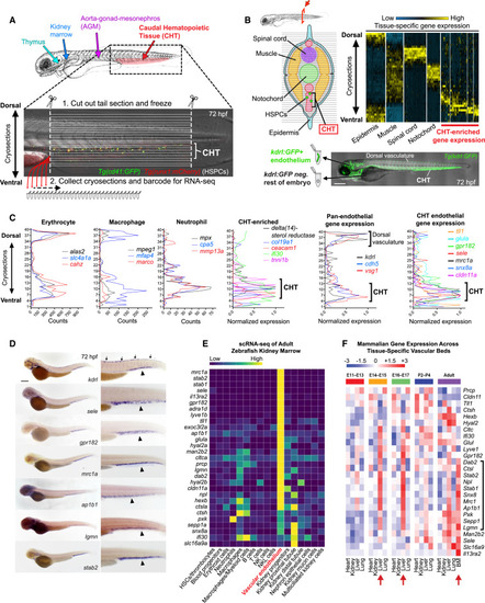
An endothelial gene expression signature unique to HSPC niches (A) Schematic diagram illustrates the hematopoietic tissues of the zebrafish embryo (top) and the sectioning strategy used to perform RNA tomography (tomo-seq) on the CHT (bottom; double transgenic embryo carrying the HSPC markers cd41:GFP and runx1:mCherry is shown). (B) Cross-section schematic (upper left) and hierarchical clustering heatmap (upper right) reveal clusters of gene expression that correspond to distinct tissues along the dorsal-ventral axis of the zebrafish tail. Schematic at bottom depicts strategy using kdrl:GFP transgenic embryos and FACS to isolate ECs from whole embryos for analysis by RNA-seq. (C) Graphs show tomo-seq expression traces for individual tissue-specific genes. (D) Images show whole-mount in situ hybridization (WISH) for the pan-endothelial gene kdrl (top panel) and CHT EC-enriched genes identified by tomo-seq and tissue-specific RNA-seq (bottom panels). Arrows point to expression in dorsal vasculature and arrowheads point to expression in the CHT. (E) Heatmap shows the expression of the 29 CHT EC genes in the different cell populations that comprise the adult zebrafish kidney marrow. Spectral scale reports normalized expression. (F) Heatmap shows the expression of orthologs of the zebrafish CHT EC genes in ECs from different organs of the mouse at different stages of development and postnatal transition to adulthood. Red arrows denote hematopoietic tissues at the respective stage of development. Black bracket denotes genes enriched in fetal liver ECs at the E14–17 stages and then later in the adult bone marrow. Spectral scales report Z scores. BM, bone marrow. Scale bars represent 250 μm in this and all subsequent figures unless noted otherwise. |
Reprinted from Developmental Cell, 58(12), Hagedorn, E.J., Perlin, J.R., Freeman, R.J., Wattrus, S.J., Han, T., Mao, C., Kim, J.W., Fernández-Maestre, I., Daily, M.L., D'Amato, C., Fairchild, M.J., Riquelme, R., Li, B., Ragoonanan, D.A.V.E., Enkhbayar, K., Henault, E.L., Wang, H.G., Redfield, S.E., Collins, S.H., Lichtig, A., Yang, S., Zhou, Y., Kunar, B., Gomez-Salinero, J.M., Dinh, T.T., Pan, J., Holler, K., Feldman, H.A., Butcher, E.C., van Oudenaarden, A., Rafii, S., Junker, J.P., Zon, L.I., Transcription factor induction of vascular blood stem cell niches in vivo, 1037-1051.e4, Copyright (2023) with permission from Elsevier. Full text @ Dev. Cell