
Fig. 4
- ID
- ZDB-FIG-231107-43
- Publication
- Windell et al., 2023 - The influence of size and surface chemistry on the bioavailability, tissue distribution and toxicity of gold nanoparticles in zebrafish (Danio rerio)
- Other Figures
- All Figure Page
- Back to All Figure Page
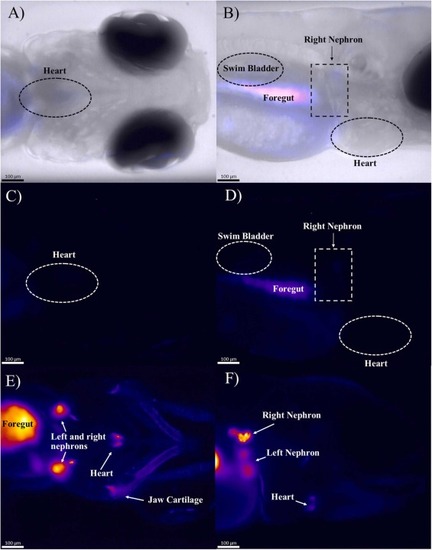
SPIM images of Casper zebrafish embryo-larvae at 20 ×. Non-exposed controls (A–D) and zebrafish embryo-larvae exposed to 2 mg/L 80 nm AuNPs (E–F). Brightfield imaging of the control provided a reference for specific organs with autofluorescence overlaid (A–B). The non-exposed, control embryo-larvae showed minimal fluorescence in both ventral and dorsal view (C & D respectively). The exposed embryo-larvae showed AuNP accumulation in the heart and the jaw cartilage (E). In the profile view (F), fluorescence can be seen in the jaw, in the right nephron and (blurred image) in the left nephron. Autofluorescence is visible in the gut (D) but the fluorescent signal is significantly higher in the AuNP exposed zebrafish (E & F). |