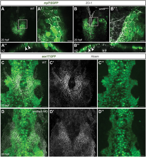
Wnt9a/b control cardiac cone formation and leftward jogging independently of epithelial cell polarity or endoderm. (A-B″) Maximum projections of confocal z-scans with ventral views (A,A′ and B,B′) or x-z section views (A″,B″) of the zebrafish heart field. Maximum projection of a 7 µm deep z-stack showing subapical tight junctional ZO-1 staining within the myocardium at 20 hpf (A′). x-z views (A″) show the correct subapical localization of tight junctions between cardiomyocytes (white arrowheads; n=9/9 embryos analyzed). In wnt9DKO mutants, apico-basal cell polarity is intact, as indicated by a maximum projection of confocal z-scans (B) or of a 7 µm deep z-stack (B′), which reveals intact tight junctions. x-z view (B″) of the wnt9DKO heart field which shows intact tight junctions within the myocardium (white arrowheads; n=12/12 embryos analyzed). (C-D″) Maximum projections of confocal z-scans with ventral views of cardiomyocyte progenitor cells during cardiac cone formation and of the underlying endoderm. In the wildtype (C-C″), the endoderm is present at the midline while the cardiac cone is completing its fusion. In wnt9a/b double morphants (D-D″), the endoderm is present at the embryonic midline, whereas the anterior portion of the cardiac cone does not migrate towards the embryonic midline and fails to fuse into the heart cone (n=15/15 embryos analyzed). Scale bars: 30 µm (A,B,C-D″); 10 µm (A′,B′).
|
