
Fig. 5
- ID
- ZDB-FIG-230717-130
- Publication
- Nakajima et al., 2023 - Endoderm-derived islet1-expressing cells differentiate into endothelial cells to function as the vascular HSPC niche in zebrafish
- Other Figures
- All Figure Page
- Back to All Figure Page
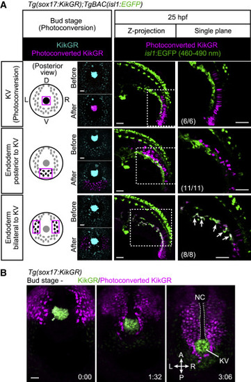
Figure 5. The isl1+ endothelial progenitors derive from endodermal cells (A) Tg(sox17:KikGR);TgBAC(isl1:EGFP) embryos at bud stage (10 hpf) photoconverted in the KV (upper) or in endodermal cells posterior to the KV (middle) or lateral to the KV (lower) as indicated on the left. Left, posterior views at bud stage; middle and right, lateral views at 25 hpf. Arrows point to photoconverted cells expressing isl1:EGFP. EGFP fluorescence can be separated by 460–490 nm range of emission filter as in Figures S4J and S4K. Left and middle, projection views; right, single confocal planes. The number of embryos analyzed in each case is indicated in the right panels. (B) Time-sequential confocal images of a Tg(sox17:KikGR) embryo (from the bud stage) just after KikGR was photoconverted in endodermal cells located lateral to the KV. Posterior view. Scale bars, 50 μm. KV, Kupffer’s vesicle; D, dorsal; V, ventral; L, left; R, right. See also Figure S4 and Videos S5 and S6. |
Reprinted from Developmental Cell, 58(3), Nakajima, H., Ishikawa, H., Yamamoto, T., Chiba, A., Fukui, H., Sako, K., Fukumoto, M., Mattonet, K., Kwon, H.B., Hui, S.P., Dobreva, G.D., Kikuchi, K., Helker, C.S.M., Stainier, D.Y.R., Mochizuki, N., Endoderm-derived islet1-expressing cells differentiate into endothelial cells to function as the vascular HSPC niche in zebrafish, 224-238.e7, Copyright (2023) with permission from Elsevier. Full text @ Dev. Cell