
Fig. 8
- ID
- ZDB-FIG-230714-36
- Publication
- Deshmukh et al., 2023 - Antiangiogenic potential of Lepista nuda extract suppressing MAPK/p38 signaling-mediated developmental angiogenesis in zebrafish and HUVECs
- Other Figures
- All Figure Page
- Back to All Figure Page
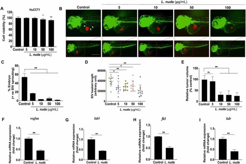
Fig. 8. Representative and quantitative results of the in vivo zebrafish xenograft angiogenesis assay. (A) Cell viability evaluation showed slightly significant cytotoxicity of L. nuda extract (0, 5, 10, 50, and 100 µg/mL) for 24 h. (B) Zebrafish xenograft assays showed that L. nuda extract-treated HuCCT1 cells inhibited SIV sprouting in Tg (fli1: EGFP) zebrafish embryos at 24 hpi. Red fluorescence: DiI-stained HuCCT1 cells; green fluorescence: the vascular network of the zebrafish embryo. White arrows indicate the location of SIV sprouting. White arrow: SIV sprouting. (C-D) Quantitative results of angiogenesis in zebrafish embryos showing SIV inhibition (n = 50) and inhibition of SIV basket length. (E) Quantitative results of in vivo tumor proliferation show depletion of tumor mass following L. nuda extract treatment. (F-I) qPCR results show that L. nuda extract treatment inhibits the mRNA expression of vegfaa, kdrl, flt1and, kdr. Scale bar = 100 µm. * p < 0.05, * * p < 0.001. |