Fig. 4
- ID
- ZDB-FIG-230712-32
- Publication
- Espinosa-Medina et al., 2022 - TEMPO enables sequential genetic labeling and manipulation of vertebrate cell lineages
- Other Figures
- All Figure Page
- Back to All Figure Page
|
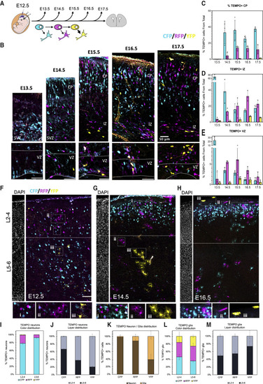
TEMPO connects sequential cell birth date and layer position in the mouse brain (A) In utero electroporation (IUE) of ubiquitous TEMPO constructs into E12.5 mouse brains and analysis at subsequent developmental stages. (B) Representative confocal images of mouse cortex at different stages following electroporation of TEMPO at E12.5. VZ, ventricular zone. SVZ, subventricular zone. IZ, intermediate zone. CP, cortical plate. Scale bars, 50 μm. (C–E) Distribution of TEMPO reporters in VZ, IZ, and CP along time. Plots represent the mean percentage of fluorescent cells from the total of cells in that brain section ± SEM, 3 independent experiments. (F–H) Postnatal P10 brain sections from mice electroporated with TEMPO constructs at stages E12.5–E16.5. DAPI labeling (separated adjacent panels) shows the limit between upper (L2-4) and lower (L5-6) cortical layers (dashed line). Insets highlight representative examples of TEMPO+ neurons (i) and astrocytes (ii and iii). (I) Percentage of neurons expressing each TEMPO reporter in L2-4 or L5-6. (J) Layer distribution of TEMPO+ neurons reveals an “inside-out” pattern. (K) Neuron and glia ratio for each reporter. (L) Percentage of astrocytes expressing each TEMPO reporter in L2-4 or L5-6. (M) Layer distribution of TEMPO+ astrocytes for each reporter. Data are represented as percent ± SEM. n = 3, Scale bars (F–H) 100 μm. |

