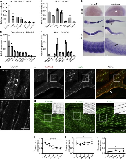
Cavin4 is associated with T-tubules of developing muscle fibers. (A and B) qRT-PCR of caveola-associated genes (relative to 36B4) in 6-mo-old adult WT mouse skeletal muscle (A) and heart tissue (B; mean± SD; n = 3 each for muscle and heart). Reactions were performed in the following groups: Cav1 and Cavin1; Cav2 and Cav3; Cavin2, Cavin3, and Cavin4. (C and D) qRT-PCR of caveola-associated genes (relative to β-actin) in 10-mo-old WT adult zebrafish trunk muscle (C) and heart (D; n = 3 for trunk muscle, n = 3 pooled samples for heart tissue). For A–D, muscle genes are shown in order of decreasing expression; heart genes are shown in the same order as for muscle samples. (E) Whole-mount ISH for cavin4a and cavin4b in 10- to 14-somite (S) and 48-hpf WT zebrafish embryos. Cavin4b expression in adaxial cells (white arrowheads), pectoral fin buds (arrow), and somite boundaries (black arrowheads) is highlighted. Images for 48 hpf shown anterior to left in both dorsal and lateral view; bottom panel shows a magnification of boxed areas. Scale bars: 200 µm. (F) Confocal images of CAVIN4 in mouse skeletal muscle before birth (16.5 dpc) and 3 d after birth (3d). Arrowheads indicate internal labeling. Scale bars: 10 µm. (G) Confocal images of CAVIN4 and CAV3 in C2C12 myotubes (arrow, sarcolemmal staining). Insets show a magnification of boxed area. Scale bars: 20 µm (inset, 10 µm). (H) Clover-tagged Cavin4a, Cavin4b, and Cavin1a in muscle fibers of 3-dpf transgenic zebrafish embryos. Inverted images = magnification of boxed area. Scale bars: 20 µm (inset, 10 µm). (I–K) Ratio of T-tubule to sarcolemmal fluorescence intensity for Cavin4a (I), Cavin4b (J), and Cavin1a (K). n = 9 muscle fibers from three embryos per line for each developmental stage (one-way ANOVA with multiple-comparison Tukey’s test). ****, P ≤ 0.0001; *, P ≤ 0.05. Error bars represent mean ± SD.
|
