
Fig. 3
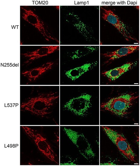
Fibroblasts heterozygous for KIF5B variants show an aberrant morphology of lysosomes and their anomalous pericellular aggregation. Confocal imaging shows morphology and distribution of lysosomes, which are enlarged and more numerous in patients’ cells compared to what is observed in control cells. Their distribution is principally in cell periphery (see also Supplementary Material, Fig. S4). In the same panels staining for mitochondria show no grossly altered distribution and morphology of the mitochondrial network. Scale bar is 5 μm. In these experiments for immunofluorescence analyses cells were stained using antibodies against Lamp1 (lysosome marker, green), TOM20 (mitochondria marker, red) and DAPI (DNA marker, blue). |