|
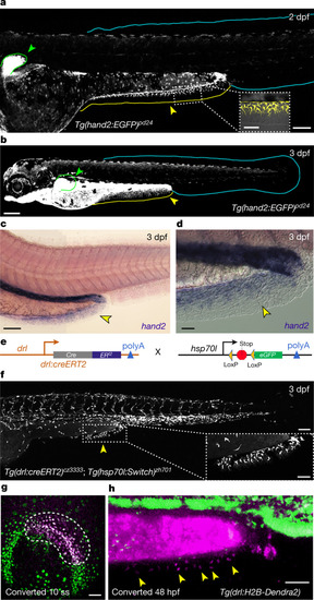
The PAFF is an LPM-derived median fin fold. a,b, Confocal images of Tg(hand2:EGFP) embryos at 2 dpf (a) and 3 dpf (b) showing eGFP labelling of the mesenchyme of the pectoral (green outline and arrowheads) and PAFFs (yellow outline and arrowheads, and magnified in inset) but not the caudal fin fold (cyan outline). c,d, In situ hybridization of hand2 at 3 dpf shows fin expression of hand2 only in the PAFF (yellow arrowheads (c), and higher magnification with Nomarski optics indicates expression in the mesenchyme (d). e, Schematic of the LPM lineage tracing transgenes. f, Lineage tracing of LPM using transgenics depicted in (e) following 4-OHT treatment and heat shock before imaging shows that PAFF mesenchyme is derived from the LPM (yellow arrowhead and magnified in inset). g,h, Ventral (g) and lateral (h) confocal images of the drl:H2B-Dendra2 transgenic line at the 10-somite stage (10 ss) (g) and 48 hpf (h) following ultra-violet laser photoconversion in the region of the LPM outlined in (g). h, Photoconverted PAFF mesenchyme is indicated by yellow arrowheads. Scale bars, 100 µm (a,c,f); 50 µm (a (inset),f (inset),g,h); 200 µm (b); 20 µm (d).
|