
Fig 2
- ID
- ZDB-FIG-230519-29
- Publication
- Viana et al., 2023 - Hiding in the yolk: A unique feature of Legionella pneumophila infection of zebrafish
- Other Figures
- All Figure Page
- Back to All Figure Page
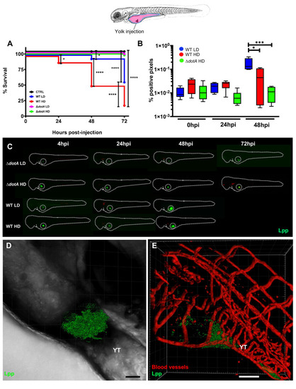
Bloodstream L. pneumophila establish a proliferative niche in the yolk causing a persistent local infection.
A) Survival curves (two independent experiments pooled) of zebrafish larvae injected into the yolk with WT-GFP Low Dose (WT LD) (blue curve, n = 48) or High Dose (HD) (red curve, n = 48), or with ΔdotA-GFP Low Dose (ΔdotA LD) (magenta curve, n = 48) or High Dose (ΔdotA HD) (green curve, n = 48), and incubated at 28°C. Non-injected fish (CTRL, black curve; n = 48). The scheme of the zebrafish larvae has been adapted from [35] and has been previously modified from [87]. B) Bacterial burden evaluation by quantification of % of fluorescent pixel counts on individual injected larvae followed over time by 0 to 48 hpi. Each larva was imaged daily, and images were analysed with Fiji for bacterial burden evaluation. One experiment plotted, 6 larvae per condition. P < 0.05 was considered statistically significant (symbols: **** P < 0.0001; ***P < 0.001; **P < 0.01; *P < 0.05. No symbol on graphs means that not statistically differences were observed). C) Representative images of L. pneumophila localization, determined by live imaging using a fluorescence stereomicroscope, of zebrafish larvae infected with a LD or a HD of WT-GFP, or a LD or a HD of ΔdotA-GFP. Individual infected larvae were followed over time by live imaging at 4h, 24h, 48h, and 72 hpi. GFP fluorescence of the injected bacteria is shown. Dotted circle highlights GFP bacteria in the yolk. D) Representative maximum intensity projection of confocal acquisition of a 72 hpi zebrafish larva injected in the bloodstream with HD of WT-GFP, mounted laterally and live imaged using high resolution confocal fluorescence microscope, showing bacteria growing in the yolk and yolk tube (YT) region. The x-y-z raw data were post treated with the LEICA lighting application for reducing noise and processed with Imaris for 3D volume rendering. Related to S1 Movie. E) Imaris 3D reconstruction and volume rendering of the L. pneumophila growth (GFP labelling) in the yolk of kdrl:ras-mCherry (red vessels) infected larva at 72hpi, showed laterally. Overlay of GFP and mCherry is shown; BF is shown to help to visualize the yolk region and host anatomy. YT Related to S2 Movie. Scale bar = 50mm. |