
FIGURE
Fig. 3
- ID
- ZDB-FIG-230503-12
- Publication
- Siebert et al., 2022 - Rhabdomyosarcoma xenotransplants in zebrafish embryos
- Other Figures
- All Figure Page
- Back to All Figure Page
Fig. 3
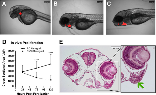
Development of RMS xenografts, derived from RD cells, is depicted at (A) 24 hpf, (B) 72 hpf and (C) 120 hpf. (D) The development of RD and Rh30 cells xenotransplants was observed until 120 hpf. The SCSA of RD tumors increased, whereas the SCSA of Rh30 tumors decreased during the observation period. (E) The head of a xenotransplanted zebrafish embryo was sectioned and stained with hematoxylin and eosin. The arrow marks a nest of tumor cells |
Expression Data
Expression Detail
Antibody Labeling
Phenotype Data
Phenotype Detail
Acknowledgments
This image is the copyrighted work of the attributed author or publisher, and
ZFIN has permission only to display this image to its users.
Additional permissions should be obtained from the applicable author or publisher of the image.
Full text @ Pediatr Blood Cancer