
Fig. 4
- ID
- ZDB-FIG-230327-4
- Publication
- Akbari et al., 2022 - Whole-brain optical access in a small adult vertebrate with two- and three-photon microscopy
- Other Figures
- All Figure Page
- Back to All Figure Page
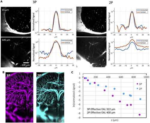
Characterization of 2PM and 3PM (both with 1280 nm excitation) images through shallow and deep regions of the adult D. dracula brain (A) Brightness comparison of horizontal and vertical blood vessels for 3PM and 2PM images at various depths inside the brain. In each line profile plot, the values are normalized to the maximum brightness of the horizontal blood vessel. (B) Maximum projection of a column containing the deepest part of the brain. (C) Characterization of effective attenuation length inside the brain for 2PM and 3PM excitation wavelengths as described in the STAR Methods section. EAL is measured to be 400 μm with 2PM images and 322 μm with 3PM. |