
FIGURE
Fig. 3
- ID
- ZDB-FIG-230327-38
- Publication
- Shin et al., 2022 - Three-dimensional fluorescence microscopy through virtual refocusing using a recursive light propagation network
- Other Figures
- All Figure Page
- Back to All Figure Page
Fig. 3
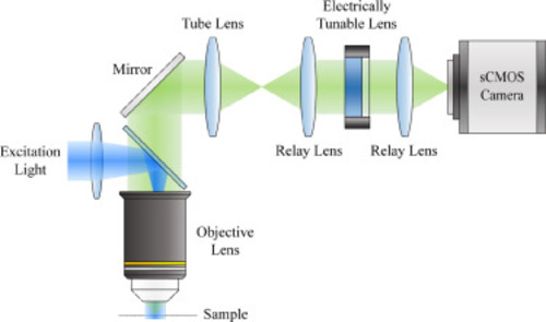
Fig. 3. Schematic of the custom-designed microscope used for imaging larval zebrafish brain. An electrically tunable lens (ETL) is conjugated to the back pupil plane of the objective lens. Rapid axial scanning can be performed by electrically modulating the focal length of the ETL. |
Expression Data
Expression Detail
Antibody Labeling
Phenotype Data
Phenotype Detail
Acknowledgments
This image is the copyrighted work of the attributed author or publisher, and
ZFIN has permission only to display this image to its users.
Additional permissions should be obtained from the applicable author or publisher of the image.
Reprinted from Medical image analysis, 82, Shin, C., Ryu, H., Cho, E.S., Han, S., Lee, K.H., Kim, C.H., Yoon, Y.G., Three-dimensional fluorescence microscopy through virtual refocusing using a recursive light propagation network, 102600, Copyright (2022) with permission from Elsevier. Full text @ Med. Image Anal.