
Fig. 2
- ID
- ZDB-FIG-230324-2
- Publication
- Xie et al., 2022 - Cilia regulate meiotic recombination in zebrafish
- Other Figures
- All Figure Page
- Back to All Figure Page
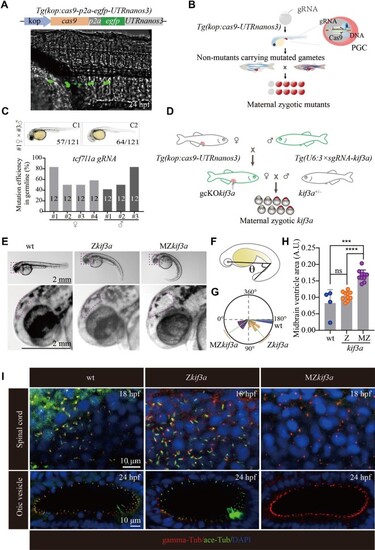
Generation of MZkif3a mutants via germ cell-specific expression of Cas9. (A) Bright field image showing a 24 hpf Tg(kop:cas9-p2a-egfp-UTRnanos3) zebrafish embryo with EGFP fluorescence in the PGCs. Top: diagram of the construct for making this transgene. (B) Schematic workflow showing process of generating MZ mutants using PGC-specific Cas9-expressing Tg(kop:cas9-UTRnanos3) embryos. (C) Mutation efficiencies of gametes and the phenotypes of offspring of tcf7l1a sgRNA-injected Tg(kop:cas9-UTRnanos3) fish. C1 shows the wild type-like phenotype, and C2 shows complete loss of eyes. The mutation efficiencies were calculated by the number of mutated heterozygotic embryos from crossing between F0 and wild type fish. A total of 12 embryos were genotyped from each cross and the mutation efficiency was calculated. (D) Schematic diagram showing the strategy for generating germ cell-specific knockout mutants of kif3a (gcKOkif3a). (E) External phenotypes of 48 hpf Zkif3a and MZkif3a mutants. The enlarged boxes are shown in the bottom, and dots outline the midbrain ventricles. (F and G) Statistical analysis showing the increased body curvature severity as demonstrated by the reduced angles of MZkif3a mutants. (H) Bar graph showing relative size of midbrain ventricles in different mutants as indicated. A.U., arbitrary unit. (I) Confocal images showing cilia in the spinal cord and otic vesicle of wild type, Zkif3a and MZkif3a mutants as indicated. Cilia were labelled with acetylated tubulin in green and the basal bodies were stained with gamma tubulin in red. Nuclei were counterstained with DAPI. ***P < 0.001; ****P < 0.0001. ns, not significant; hpf, hours post-fertilization; wt, wild type. |