FIGURE

Figure 3

ID
ZDB-FIG-230228-104
Publication
Lu et al., 2023 - A CRISPR-Cas9-mediated F0 screen to identify pro-regenerative genes in the zebrafish retinal pigment epithelium
Other Figures
All Figure Page
Back to All Figure Page
Figure 3

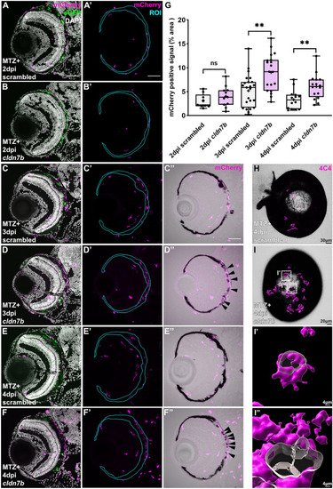
cldn7b F0 larvae show retention of macrophages/microglia and impaired clearance of pigment debris during RPE regeneration. (AF) Representative immunofluorescence images of mCherry signal in ablated (MTZ +) scrambled and cldn7b F0 larvae at (A,B) 2dpi, (C,D) 3dpi, and (E,F) 4dpi. (A’F’) Single-channel images of mCherry signal in RPE ROIs. (C’’F’’) Overlay of mCherry and bright-field channels. Nuclei (white), eGFP (green), mCherry (magenta). (G) Box plots showing significantly more mCherry+ macrophage/microglia in the RPE layer of cldn7b F0 larvae at 3dpi and 4dpi, compared to scrambled controls. (H,I) Overlaid images of 3D-rendered 4C4+ isosurfaces (magenta) with brightfield channels from ablated scrambled and cldn7b knockout whole-mount eyes. 4C4+ macrophages/microglia (magenta) surfaces were 3D-rendered in Imaris. (I’, I’’) Zoom-in of (I; white boxes) cldn7b F0 whole-mount eye showing 4C4+ cells engulfing pigment debris. (AF) Scale bars = 50 μm. (HI) Scale bars were indicated on the images. **p value ≤ 0.01; ns not significant. Exact p values, numbers of independent experiments (N), and numbers of biological replicates (n) can be found in Table S3. Abbreviations as follows: MTZ Metronidazole; dpi Days post-injury; ROI Region of interest.

Expression Data

Expression Detail
Antibody Labeling
Phenotype Data

Phenotype Detail
Acknowledgments
This image is the copyrighted work of the attributed author or publisher, and ZFIN has permission only to display this image to its users. Additional permissions should be obtained from the applicable author or publisher of the image. Full text @ Sci. Rep.