
FIGURE
Fig. 5
- ID
- ZDB-FIG-230204-32
- Publication
- Brown et al., 2021 - A novel gene trap line for visualization and manipulation of erbb3b+ neural crest and glial cells in zebrafish
- Other Figures
- All Figure Page
- Back to All Figure Page
Fig. 5
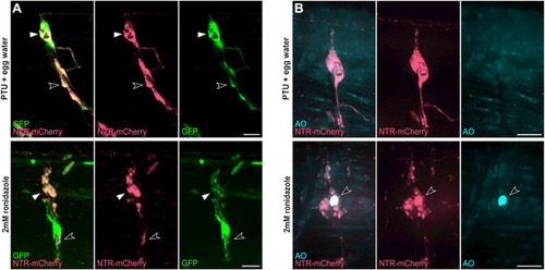
Fig. 5. Cell specific ablation using the erbb3b-driven transgene (A) Tg(UAS:NTR-mCherry);gSAIzGFFD37A larvae at 120 hpf after 24 h treatment of phenyl-thiourea (PTU) and egg water (Top) or 2 mM ronidazole (Bottom) with labeled satellite glia (white arrowheads) and Schwann cells (open arrowheads) in dorsal root ganglia (DRG). (B) Acridine orange (AO) staining of apoptotic cells (arrowhead) in Tg(UAS:NTR-mCherry);gSAIzGFFD37A (GFP-negative) larvae at 120 hpf after 24 h treatment of PTU and egg water (Top) or 2 mM ronidazole (Bottom). Scale bars, 20 μm unless otherwise noted. |
Expression Data
Expression Detail
Antibody Labeling
Phenotype Data
Phenotype Detail
Acknowledgments
This image is the copyrighted work of the attributed author or publisher, and
ZFIN has permission only to display this image to its users.
Additional permissions should be obtained from the applicable author or publisher of the image.
Reprinted from Developmental Biology, 482, Brown, E.A., Kawakami, K., Kucenas, S., A novel gene trap line for visualization and manipulation of erbb3b+ neural crest and glial cells in zebrafish, 114-123, Copyright (2021) with permission from Elsevier. Full text @ Dev. Biol.