
Fig. 9
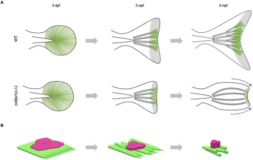
Fig. 9. The inferred actinotrichia function in zebrafish tail fin formation. (A) Growth of fins in WT (upper images) and col9a1c(-/-) (lower images) fish. In WT, actinotrichia are always dense at the tip, whereas in col9a1c(-/-), the number of actinotrichia decreases and their orientation becomes abnormal as the fish gets older. The fin gradually retracts along the DV axis, and the fin-ray also bends toward the center (blue dot arrows). In addition, there is little branching of the fin-ray. The green lines indicate the actinotrichia and the white bars indicate the fin-rays. The area of the actinotrichia at the tip of the fin is shown in grey. (B) Schematic diagram of the non-spread epidermal cells due to the disorderly actinotrichia arrangement in col9a1c(-/-). Each step corresponds to the fin growth in A. |
Reprinted from Developmental Biology, 481, Nakagawa, H., Kuroda, J., Aramaki, T., Kondo, S., Mechanical role of actinotrichia in shaping the caudal fin of zebrafish, 52-63, Copyright (2021) with permission from Elsevier. Full text @ Dev. Biol.