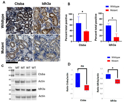
Immunohistochemical and Western blot detection of selected proteins in kidneys from WT and M ZF reveals protein expression disturbances in mitochondria and lysosomes. (A) Representative IHC staining specific for mitochondrial marker isocitrate dehydrogenase (NAD(+))3 alpha (Idh3a) and lysosomal marker cathepsin B (Ctsb) in kidney tissue sections from WT and M ZF (upper right and upper left panels, and lower right and lower left panels, respectively; (B) Quantification of immunohistochemical staining of sections from WT and M ZF kidneys. Signal intensity is significantly higher in WT than in M for both proteins. (C) Representative immunoblots of Ctsb and Idh3a in kidney tissue section from WT and M ZF; (D) Quantification of immunoblots from WT and M ZF kidneys. (Mann–Whitney test U * p < 0.05). Wildtype (WT), mutant (M). ns: not significant. Scale bar (bottom left corner, in black) = 100 µm.