
FIGURE
Fig. 5
- ID
- ZDB-FIG-221219-12
- Publication
- Huisman et al., 2021 - Meningeal lymphatic endothelial cells fulfill scavenger endothelial cell function and cooperate with microglia in waste removal from the brain
- Other Figures
- All Figure Page
- Back to All Figure Page
Fig. 5
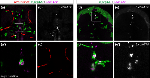
Microglia but not BLECs internalize CFP-labeled Escherichia coli from the brain parenchyma. (a-c) Maximum projection of the head region (dorsal view) of a lyve1:DsRed; mpeg:GFP double transgenic embryo that was injected with CFP-labeled E. coli at 5dpf. (a') Single confocal slice of the boxed area in a, showing uptake of E. coli by a GFP-positive macrophage. (d-e') Partial projection of a mpeg:GFP positive embryo that was injected with CFP-labeled E. coli. The box indicates the area of zoom-in in (d′) and (e'). Note that the green signal marked by an asterisk in (d) represents auto-fluorescence of the embryo
|
Expression Data
| Genes: | |
|---|---|
| Fish: | |
| Condition: | |
| Anatomical Terms: | |
| Stage: | Day 5 |
Expression Detail
Antibody Labeling
Phenotype Data
| Fish: | |
|---|---|
| Condition: | |
| Observed In: | |
| Stage: | Day 5 |
Phenotype Detail
Acknowledgments
This image is the copyrighted work of the attributed author or publisher, and
ZFIN has permission only to display this image to its users.
Additional permissions should be obtained from the applicable author or publisher of the image.
Full text @ Glia