|
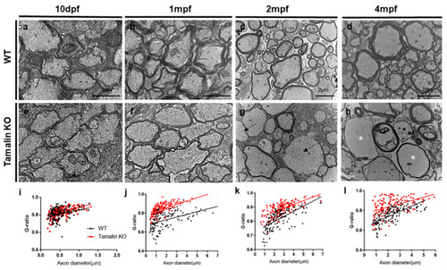
Tamalin KO causes the hypomyelination of axons in the zebrafish spinal cord. Transmission electron microscopy (TEM) images reveal transverse sections through the spinal cord of wildtype or tamalin KO zebrafish at 10 dpf (a,e), 1 mpf (b,f), 2 mpf (c,g), and 4 mpf (d,h). Black arrows indicate unmyelinated axons (g), and asterisks indicate disorganized watery axons (h). (i–l) The g-ratio of myelinated axons in the spinal cord of wildtype and tamalin KO zebrafish. Unpaired t test was used to compare means from each animal. Each g-ratio has been obtained from 100 myelinated axons in eight sections of four zebrafish each (i: p = 0.0696, j–l: p < 0.0001). Scale bars: (a,e): 1 μm, (b–h): 2 μm. KO, knockout; dpf, days post fertilization; and mpf; months post fertilization.
|