
Fig. 4
- ID
- ZDB-FIG-220928-10
- Publication
- Jing et al., 2021 - A BMP4-p38 MAPK signaling axis controls ISL1 protein stability and activity during cardiogenesis
- Other Figures
- All Figure Page
- Back to All Figure Page
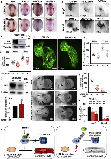
Figure 4. Inhibition of p38 results in ISL1 degradation in vivo and in defects in cardiac morphogenesis and function (A) In situ hybridization of zebrafish embryos at the ten somite stage for isl1, mef2ca, mef2cb, and nkx2.5 expression. The embryos were treated either with DMSO or SB202190 starting from the three somite stage. Scale bar, 0.2 mm. (B) WB analysis of ISL1/2 in 72 hpf zebrafish embryos treated either with DMSO or p38 inhibitor at 24 hpf (at least 20 embryos per sample). (C) Heart beats per minute at 48 and 72 hpf of zebrafish embryos treated either with DMSO or p38 inhibitor at 24 hpf. Error bars represent SEM of four clutches. The numbers of embryos used per condition are indicated in Figure S3F. (D) Representative confocal images of control and p38 inhibitor-treated Tg(myl7:EGFP-HsHRAS)s883 embryos at 72 hpf. Scale bar, 50 μm. (E) Quantification of ventricular CM numbers at 48 and 72 hpf of zebrafish embryos treated either with DMSO or p38 inhibitor at 24 hpf. DMSO, n = 15; 50 μM SB202190, n = 15, from 3 clutches. (F) Representative in situ hybridization for ltbp3 (top) or bmp4 (bottom) of zebrafish embryos at 48 hpf treated either with DMSO or p38 inhibitor at 24 hpf as well as of isl2b−/− or isl1−/− embryos. The arrow points to the bmp4 expression at the venous pole lost upon p38 inhibitor treatment and in isl1−/− embryos. Scale bar, 0.1 mm. (G) WB analysis for ISL1 of protein extracts from E8.5 mouse embryos treated either with DMSO or 50 μM SB202190 for 24 h in a whole-embryo ex vivo culture (at least five embryos per sample). (H) Relative Isl1 mRNA expression in ex-vivo-cultured mouse embryos treated either with DMSO or 50 μM SB202190. (I) Representative right lateral views of the hearts of embryos treated either with DMSO (n = 27) or 20 μM (n = 9) and 50 μM SB202190 (n = 26) at E8.5 for 24 h. Scale bar, 0.2 mm. (J) Heart beats per minute of ex-vivo-cultured mouse embryos treated either with DMSO or SB202190 (n = 8). (K) Relative mRNA expression of SAN lineage markers Hcn4 and Shox2 in ex-vivo-cultured mouse embryos treated either with DMSO or 20 and 50 μM SB202190 starting at E8.5 for 24 h (n = 6). (L) Model of the role of BMP-p38 MAPK signaling axis in ISL1-driven cardiogenesis. In WT CPCs, BMP signaling activates p38 leading to ISL1 phosphorylation at S269 thereby preventing ISL1 degradation and ensuring its transcriptional activity during cardiogenesis. Interfering with the BMP-p38 MAPK signaling axis leads to degradation of ISL1 by the proteasome, resulting in defects in CM differentiation and impaired SHF development. |