
Fig. 6
- ID
- ZDB-FIG-220912-117
- Publication
- Wang et al., 2022 - Tankyrase Inhibition Attenuates Cardiac Dilatation and Dysfunction in Ischemic Heart Failure
- Other Figures
- All Figure Page
- Back to All Figure Page
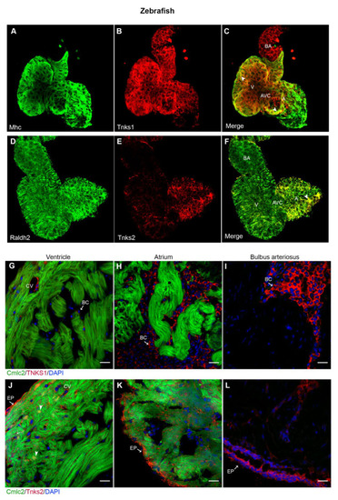
Tnks1 and Tnks2 display a distinct expression pattern in zebrafish developing and adult hearts. (A–C) Confocal images of whole-mount immunostaining for Mhc (A) and Tnks1 (B) in dissected zebrafish heart at 4 dpf. Tnks1 is co-expressed with Mhc in the ventricle (arrowhead) and atrium (arrowhead): A, atrium; V, ventricle; BA, bulbus arteriosus; AVC, atrioventricular canal. (D–F) Representative confocal images of whole-mount immunostaining for Raldh2 (D) and Tnks2 (E) in dissected zebrafish heart at 4 dpf. Tnks2 is co-expressed with Radldh2 in the epicardium (arrowhead). (G–I) Representative immunostaining of adult heart sections from transgenic zebrafish Tg(cmlc2:GFP) for Tnks1 (in red) in ventricle (G), atrium (H), and bulbus arteriosus (I). The Tnks1-positive signal is seen in the coronary vasculature (CV) and blood cell (BC) in the lumen. (J–L) Representative immunostaining of adult heart sections from transgenic zebrafish Tg(cmlc2:eGFP) for Tnks2 (in red) in ventricle (J), atrium (K), and bulbus arteriosus (L). The Tnks2-positive signal is seen in the epicardium (EP), the nucleus of the trabecular and cortical cardiomyocytes (arrowhead), and the coronary vasculature (CV). Scale bars: 20 μm. |