
Fig. 4
- ID
- ZDB-FIG-220829-233
- Publication
- Rea et al., 2022 - Gut-derived metabolites influence neurodevelopmental gene expression and Wnt signaling events in a germ-free zebrafish model
- Other Figures
- All Figure Page
- Back to All Figure Page
|
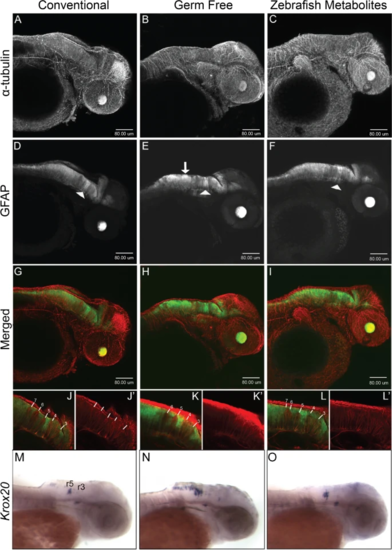
Neural development is disrupted in germ-free embryos. Confocal projection images of zebrafish embryos at 2dpf. A–C α-tubulin immunostaining. D–F GFAP:GFP fluorescence displays a non-uniform distribution in the hindbrain in germ free embryos (white arrow in E) and to some extent in ZM-treated embryos. The white arrowheads identify the GFAP tract between rhombomeres 4 and 5 which do not appear to be significantly altered in germ-free embryos. G–I Merged images of α-tubulin and GFAP to GFP. J–L Representative single-layer images of regions in the hindbrain. In conventional embryos, rhombomere tracts, 3–7 are readily identifiable by the relative absence of GFAP fluorescence. The higher intensity GFP to GFAP fluorescence between rhombomeres 4 and 5 provides a landmark for their easy identification. Note the absence of rhombomere 7 in germ-free embryos, and the seemingly merged tracts 6 and 7 in ZM-treated embryos. More examples are presented in Supplementary Figs. 5 and 6. M-O WMISH of krox20 in 2dpf embryos from CV (M), GF (N) and ZM treated (O) embryos. Rhombomeres 3 (r3) and 5 (r5) are labelled. Larvae from each treatment group were processed in parallel 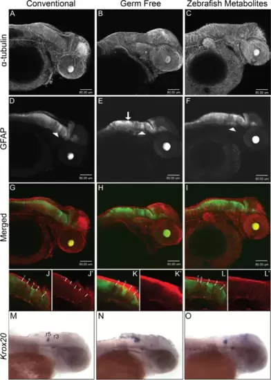
|