
FIGURE
Fig. 3
- ID
- ZDB-FIG-220809-45
- Publication
- Yáñez et al., 2021 - The organization of the zebrafish pallium from a hodological perspective
- Other Figures
- All Figure Page
- Back to All Figure Page
Fig. 3
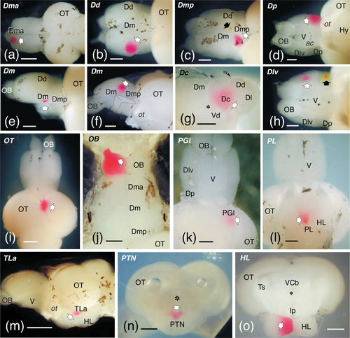
Photographs of brains showing examples of the areas labeled with DiI (outlined arrows) and DiO (black arrows) in various types of experiments. Most applications were done in toto. (a)–(c), and (j) are dorsal views. (d), (h), (k), and (l) are ventral views and (m) is a lateral view. (e) and (f) are dorsal and medial views of the same brain in which the left hemisphere was removed. Examples also include applications to transverse-sectioned en-block brains (g, n, o). In (a–f), (h), and (m), rostral is to the left; in (i–l), rostral is to the top. In (g), (n), and (o), dorsal is to the top. Scale bars: 500 μm (a, i, m); 250 μm (b–h, j–l, n–o)
|
Expression Data
Expression Detail
Antibody Labeling
Phenotype Data
Phenotype Detail
Acknowledgments
This image is the copyrighted work of the attributed author or publisher, and
ZFIN has permission only to display this image to its users.
Additional permissions should be obtained from the applicable author or publisher of the image.
Full text @ J. Comp. Neurol.