
FIGURE
Fig. 2
- ID
- ZDB-FIG-220803-28
- Publication
- Cui et al., 2022 - PARD3 gene variation as candidate cause of nonsyndromic cleft palate only
- Other Figures
- All Figure Page
- Back to All Figure Page
Fig. 2
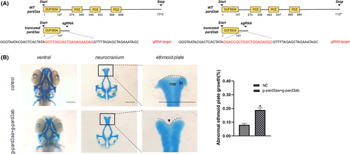
Zebrafish with pard3aa and pard3ab disruption displayed ethmoid plate patterning defects. (A) Schematic diagram of the binding and cleavage site of CRISPR/Cas9 in the coding sequence of the zebrafish PARD3 orthologs (pard3aa, pard3ab). Red = gRNA target. (B) Comparison of craniofacial structures of F0 CRISPR/Cas9-mediated mutant zebrafish and Cas9 control zebrafish. The neurocranium was dissected at 3 dpf. Compared with the larvae in the control group, the CRISPR mutant larvae had a mild dysplasia phenotype, with subtle indentations at the upper edge of the ethmoid plate or hypoplastic at the median ethmoid (arrowheads). me = median ethmoid; le = lateral ethmoid. The statistical analysis of abnormal developmental palate was performed (bars indicate the means ± SEM. Student’s t test, *p < 0.05). Scale bar = 200 μm
|
Expression Data
Expression Detail
Antibody Labeling
Phenotype Data
| Fish: | |
|---|---|
| Knockdown Reagents: | |
| Observed In: | |
| Stage: | Protruding-mouth |
Phenotype Detail
Acknowledgments
This image is the copyrighted work of the attributed author or publisher, and
ZFIN has permission only to display this image to its users.
Additional permissions should be obtained from the applicable author or publisher of the image.
Full text @ J. Cell. Mol. Med.