
Fig. 2
- ID
- ZDB-FIG-220720-21
- Publication
- Groenewoud et al., 2022 - XePhIR: the zebrafish xenograft phenotype interactive repository
- Other Figures
- All Figure Page
- Back to All Figure Page
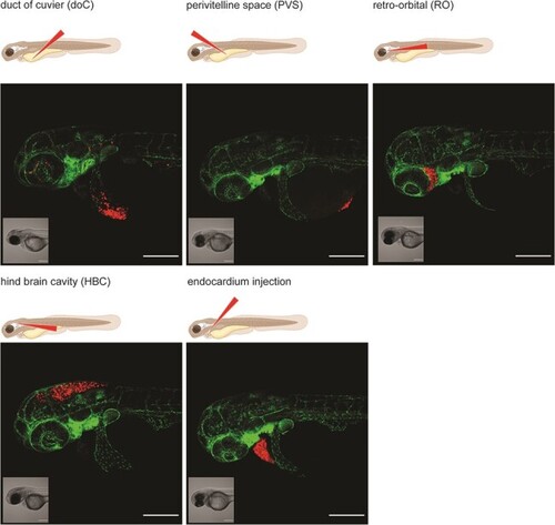
Graphical representation of common injection sites of the zebrafish xenograft model. Injection through the duct of Cuvier, a common route of injection to generate hematogenously disseminated cancer, is also described as an experimental micrometastatic model (10, 11). Perivitelline space injection, originally described as a model for the generation and assessment of angiogenesis, more recently developed as a model for the generation of primary-like tumors (6). Retro-orbital engraftment, used for the generation of orthotopic primary-like tumors derived from eye tumors, allows for the development of distant metastases (12). Hindbrain cavity injection models are used for the generation of orthotopic brain cancer models and for the generation of brain metastasis models (13). Intra-pericardial injection, used for the establishment of primary-like tumors, in an environment closed off from further blood circulation (14, 15). All larvae were injected with ∼200–300, tdTomato expressing cancer cells at 48hpf and imaged at approximately 2 h post-injection using a Zeiss LSM800 laser scanning confocal microscope with an Airyscan detector. |