Fig. 1
- ID
- ZDB-FIG-220628-124
- Publication
- Xu et al., 2022 - Reversion of MRAP2 Protein Sequence Generates a Functional Novel Pharmacological Modulator for MC4R Signaling
- Other Figures
- All Figure Page
- Back to All Figure Page
|
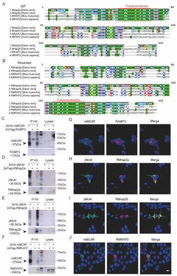
Interaction of RMRAP2 and RMrap2a/b with mMC4R and zMc4r. (A,B) Multiple sequence alignment by MUSCLE (3.8) of wild (A) and reversed (B) zebrafish Mrap2a/b, mouse MRAP2 and human MRAP2. Red frame indicates the transmembrane region. (C,G) Negative control. Mouse RAMP3 did not interact with MC4R. (D–F) Interactions of MRAP2 with MC4R proteins. Coimmunoprecipitation of 3xHA-Mc4r with 2xFlag-RMrap2a (D) or 2xFlag-RMrap2b (E) or mouse MC4R and RMRAP2 (F) in HEK293T cells. MC4R was detected with mouse anti-HA antibody; MRAP2 was detected with mouse anti-Flag antibody. IP: protein samples with anti-HA immunoprecipitation. Lysate: relevant protein samples to the samples in the IP group but without any immunoprecipitation. (G–J) Corresponding immunofluorescence of co-localization of protein complexes on the cell surface. Green indicates zMc4r or mMC4R, and red indicates RMRAP2s. Scale bar = 10 μm. mMC4R: mouse MC4R, zMc4r: zebrafish Mc4r, RMrap2a: reversed zebrafish mrap2a, RMrap2b: reversed zebrafish mrap2b, mRMRAP2: mouse MC4R. The uncropped Western blot figures were presented in Figure S2. |

