
Fig. 4
- ID
- ZDB-FIG-220628-114
- Publication
- Radhakrishnan et al., 2022 - Mice Lacking the Systemic Vitamin A Receptor RBPR2 Show Decreased Ocular Retinoids and Loss of Visual Function
- Other Figures
- All Figure Page
- Back to All Figure Page
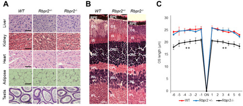
Rbpr2−/− mice on vitamin A-deficient diets show retinal phenotypes. (A,B) Representative tissue histology and staining of liver, heart, adipose, testis, retina (H&E stain), and kidney of 12-week old wild-type (WT), heterozygous Rbpr2+/−, and homozygous Rbpr2−/− (KO) mice on vitamin A-deficient diets. (C) Quantification of photoreceptor outer segment (OS) lengths from H&E sections of 12-week old WT, heterozygous Rbpr2+/−, and homozygous Rbpr2−/− (KO) mice using “spider graph” morphometry. The OS lengths from H&E sections through the optic nerve (ON; 0 μm distance from the optic nerve and starting point) were measured at 12 locations around the retina, six each in the superior and inferior hemispheres, each equally at approximately 150 μm distances. RPE, retinal pigmented epithelium; OS, outer segments; IS, inner segments; ONL, outer nuclear layer; INL, inner nuclear layer; IPL, inner plexiform layer. Scale bar = 50 µm (A); 100 µm (B). ** p < 0.005; Rbpr2−/− mice vs. controls. |