
Fig. 1
- ID
- ZDB-FIG-220622-68
- Publication
- Nguyen et al., 2022 - Dynamics of the Zebrafish Skeleton in Three Dimensions During Juvenile and Adult Development
- Other Figures
- All Figure Page
- Back to All Figure Page
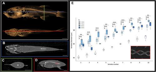
Cross sections of microCT scans visualized in 3D Slicer. (A), Surface rendering of 24 mm SL adult fish with lateral (top) and dorsal (bottom) views with coronal (blue), transverse (green), and sagittal (red) axes indicated. (B), Coronal cross section image. (C), Transverse cross section image. (D), Sagittal cross section image. (E), Quantification of vertebral canal width of 3 individual 24 mm SL adult fish; each individual differentiated with different shapes (circle, square, triangle). Inset shows higher resolution image through vertebrate (corresponding to the boxed detail in panel D). Red arrow indicates a canal from which interior width was measured. Scale bars, 2 mm. |