|
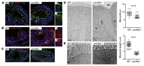
MVID features in enterocytes of <italic toggle='yes'>unc45a</italic> zebrafish mutants.(A–C) Confocal microscopy analysis of the intestinal bulb of WT and unc45a–/– mutant larvae stained for villin and pERM (A), F-actin (phalloidin) (B), and Rab11 (C) at 5 dpf. Scale bars: 20 μm. Boxes showing microvillus-like inclusions are enlarged ×4. (D and E) TEMs of thin sections of intestinal bulb of 5 dpf WT and unc45a–/– mutant larvae showing defects in the organization of the brush border. Scale bars: 2 μm (D); 0.5 μm (E). Quantitative analysis showing decrease in microvillus length and density in unc45a–/– enterocytes as compared with WT enterocytes. Data are represented as mean + SD. ****P < 0.001, t test. N nucleus; MV microvilli.
|