Fig. 5
- ID
- ZDB-FIG-220520-51
- Publication
- Brunsdon et al., 2022 - Aldh2 is a lineage-specific metabolic gatekeeper in melanocyte stem cells
- Other Figures
- All Figure Page
- Back to All Figure Page
|
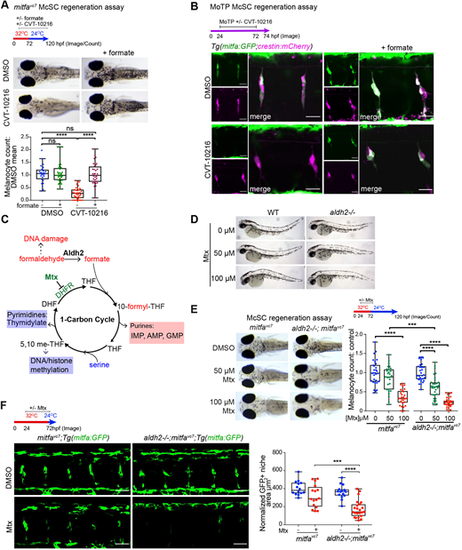
The Aldh2 metabolic reaction product, formate, promotes McSC-derived progeny. (A) Representative images of a regeneration assay where control or CVT-10216-treated embryos were supplemented with 25 mM sodium formate. ****P<0.0001; ns, not significant. Kruskal–Wallis test with Dunn's multiple comparisons. One data point per embryo; boxes indicate median and quartiles; whiskers span minimum to maximum values; three experimental replicates. (B) A MoTP assay on Tg(mitfa:GFP;crestin:mCherry) embryos treated with or without CVT-10216, and with or without 25 mM sodium formate from 24 hpf. MoTP was washed out at 72 hpf, and embryos imaged confocally at 74 hpf. Two experimental replicates, more than five embryos imaged per replicate. Scale bars: 25 µm. Single channel images of crestin:mCherry expression (magenta) and mitfa:GFP expression (green) are shown alongside merged channels. (C) Schematic of 1C metabolism and proposed function for Aldh2 formate supply through formaldehyde metabolism (based on Burgos-Barragan et al., 2017). Tetrahydrofolate (THF) combines with formate to make 10-formyl-THF, which provides two carbons to make purine nucleosides. (D) Mtx treatment has no effect on embryonic melanocytes. Zebrafish embryos (wild type and aldh2−/−) treated with or without Mtx at 24 hpf for 48 h. n=3. (E) Representative images of control and aldh2−/− mutants with or without Mtx treatment in a mitfavc7 regeneration assay. The melanocyte count at each dose was normalised to its respective DMSO control. ***P<0.0002, ****P<0.0001 (one-way ANOVA performed with Tukey's multiple comparisons test). One data point plotted per embryo; boxes indicate median and quartiles; whiskers span minimum to maximum values; three experimental replicates. (F) Confocal z-stacks of mitfa:GFP McSCs in a mitfavc7 regeneration assay, in control or aldh2−/− embryos treated with or without Mtx. Scale bars: 50 µm. Two experimental replicates; boxes indicate median and quartiles; whiskers span minimum to maximum values; more than five embryos imaged per repeat. Quantification of GFP+ niche area/somite of embryos treated with Mtx is shown. ***P<0.0002, ****P<0.0001 (one-way ANOVA with Tukey's multiple comparisons). |

