
Fig 5
- ID
- ZDB-FIG-220517-40
- Publication
- Maters et al., 2022 - Embryonic and aglomerular kidney development in the bay pipefish, Syngnathus leptorhynchus
- Other Figures
- All Figure Page
- Back to All Figure Page
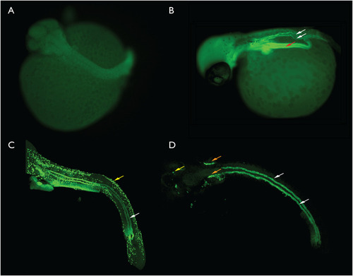
A. Late segmentation stage, no kidney is visible. B. Early pharyngula, paired kidney tubules are present and atp1a positive (white arrows). The gut tube is also fluorescent (red arrow). C. Scanning confocal generated image stack illustrating the kidney tubules (white arrow) and ionocytes (yellow arrow) in a late pharyngula embryo. D. Late pharyngula scanning confocal generated image stack. This stack was collected only in the plane of the kidney so most other labelled structures were not present. An unknown structure (orange arrows), possibly the developing gills and some ionocytes (yellow arrow) were also present in the image stack. For the purpose of scale, the eye in the early pharyngula in panel 5B is approximately 150 microns in diameter. |