|
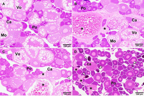
Histopathological changes in the zebrafish ovary exposed to different concentrations of BPA in water. A) Ovarian parenchyma of control zebrafish not exposed to BPA with numerous follicles at different stages of maturation, without histopathological lesions. B) Occasional presence of atretic follicles (asterisk) in ovary from zebrafish exposed to 10 μg/L of BPA. C) Presence of some atretic follicles (asterisk) and occasional presence of primordial follicles apoptosis (arrow) in ovary from zebrafish exposed to 100 μg/L of BPA. D) Multiple primary oocytes, primordial follicles apoptosis (arrows) and atretic follicles (asterisk) in ovary from zebrafish exposed to 1000 μg/L of BPA. Po-Primary oocyte; Ca-oocyte in cortical alveolus stage; Vo-vitellogenic oocyte; Mo-mature oocyte. Hematoxilin-eosine stain.
|